Understanding Your Eyes: Eye Anatomy and Function
We often take our eyes for granted. We forget how complex they are and how important our vision is for everyday life. By learning about eye anatomy and function, you will see how each small feature of the eye plays a major part. This can help you appreciate just why it is so critical to take good care of your eyes.
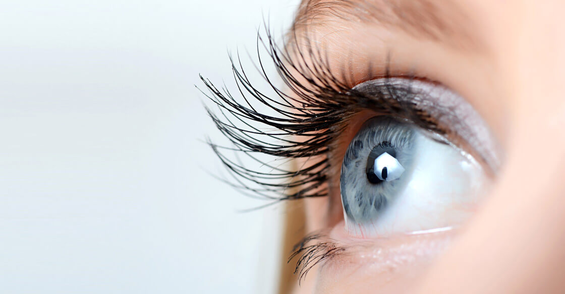
The Anatomy of the Eye
When you look at an eye, you can see some of the outer and middle layers. The outer layer is made up of the sclera (the white part of the eye) and the cornea (a clear tissue that bulges out from the sclera). The middle layer is called the choroid. Here, you find the colored iris with its opening — the pupil.
Inside the eye is the inner layer: the retina. This layer lines the back two-thirds of the eyeball. The retina is further split into two parts. The sensory retina houses nerve cells, which process visual information before it reaches the brain. The retinal pigment epithelium is behind the sensory retina and in front of the wall of the eye.
Most of the retina provides you with peripheral vision. Only the macula, a small area close to the center on the back of the eye, provides you with central vision.
Some parts of the eye do not play a direct role in vision but are still essential. For instance, the eyelids and corneal epithelium (the outer layer of the cornea) protect the eye from injury. The middle of the eye, in the space between the lens and the retina, is called the vitreous body. This is made up of a clear gel-like fluid that helps the eyeball keep its shape.
How The Eye Functions
The eye works much like a camera. The cornea is like the camera lens, allowing light to enter the eye. The light then passes through the anterior chamber, a space between the cornea and the iris. The iris controls how much light is able to reach the back of the eye by changing the size of the pupil, just like the diaphragm of a camera adjusts the aperture.
Behind the pupil is the lens, which further focuses light. The lens can control whether the eye focuses on close or far objects by contracting or relaxing.
Finally, light reaches the retina, which converts optical images into nerve impulses. This is like the image sensor of a digital camera. The many nerve fibers in the retina combine to create the optic nerve. They are joined with blood vessels at the back of the eye and travel together to the visual cortex in the brain.
Paying attention to eye health is key to maintaining normal eye anatomy and function. Receiving regular eye tests, using UV sunglasses, and following proper hygiene practices with contact lenses are all ways to protect your vision and prevent serious problems.
Schedule an Appointment