5 Things to Expect During a Digital Retinal Imaging Eye Exam

Have you been experiencing flashes of light randomly through the day while looking at an object? Or seeing floaters in front of your eyes? Are you suffering from diabetes?
If you nodded yes to any of these, then you should probably be scheduling digital retinal imaging eye exam.
Retinal imaging uses a camera-like device to take digital pictures of the most vital tissue present in your eyes, i.e., the retina. It includes photos of the retina, which senses light and perceives images, the optic disk (a spot on the retina that holds the optic nerve, which sends information to the brain), and the blood vessels.
A digital retinal imaging eye exam helps your doctor to evaluate the health of your eyes. Digital retinal imaging, vs dilation, helps to detect and treat in time certain eye diseases like diabetic retinopathy and macular degeneration.
Preparation for the Digital Retinal Imaging Eye Exam
1) An ophthalmologist will use individual drops to widen or dilate the pupils of your eyes. In about 20 minutes, your eyes will be completely dilated and ready for the examination.
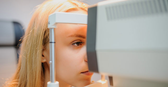
2) Next, you sit straight facing the camera.
3) To get perfect images of the retina, you’ll place your chin and forehead on a support to keep your head steady in front of the camera.
4) Your eyes will be wide open, and you’ll stare straight ahead at an object while a laser scans your eyes and takes detailed photos of the inside of your eyes. The images are uploaded to a computer so your doctor can look at them immediately.
5) The digital retinal imaging eye exam is completed in about 5 minutes.
Macular Degeneration
If your doctor suspects macular degeneration, you may probably need a fluorescein angiogram as well. For fluorescein angiography, the doctor will insert an IV needle in a vein in your arm and inject a fluorescent dye. As the dye enters your eye, it highlights the blood vessels and lets the device take some detailed photos. The photos show how the dye flows through the blood vessels in the retina and detect any blood leaks or obstructions in the blood vessels. It might identify one of the two macular degeneration conditions mentioned below-
Dry macular degeneration: A condition that occurs when the blood vessels under the retina become thin and brittle.
Wet macular degeneration: A condition that occurs when abnormal blood vessels grow under the retina causing rapid loss of vision.
Fluorescein dye may cause a metallic taste in your mouth, nausea and warm sensation. Some people may develop an allergy to the dye. Tell your doctor immediately if you experience any symptoms of lightheadedness, vomiting, and itching.

Digital Retinal Imaging Benefits and Drawbacks
Benefits
– Imaging tests allow ophthalmologists to see and identify signs of eye disorders that cannot be seen with the naked eye.
– The test is painless.
– The test images are simple for doctors to interpret.
– For future reference and comparisons, images and results are easily stored.
Your medical insurance (not vision insurance) may cover the expense of the retinal imaging, depending on the term of your policy and reason for the test.
Drawbacks
– A digital retinal imaging eye exam can’t detect the bleeding site in the retina.
– It may not be able to detect problems on the outer edges of your retina.

Eye anatomy and function can be difficult to understand. A digital retinal imaging eye exam is recommended for early detection of warning signs of retinal diseases. It can pick moles in the retina. It is the perfect test for you if you are at high risk of retinal detachment or a tear due to any of the reasons mentioned earlier.
Vision loss can be permanent! Don’t delay and get an eye exam done today!